

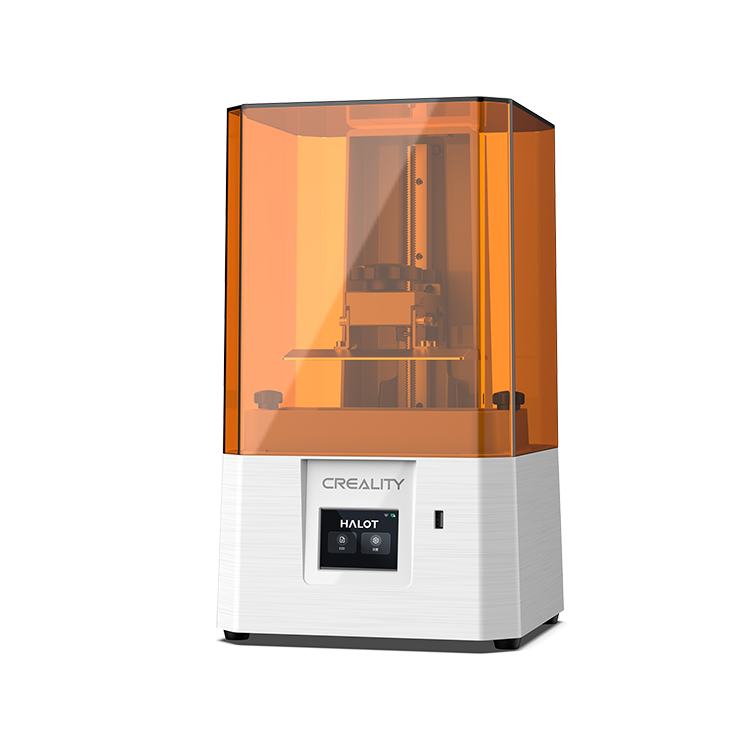
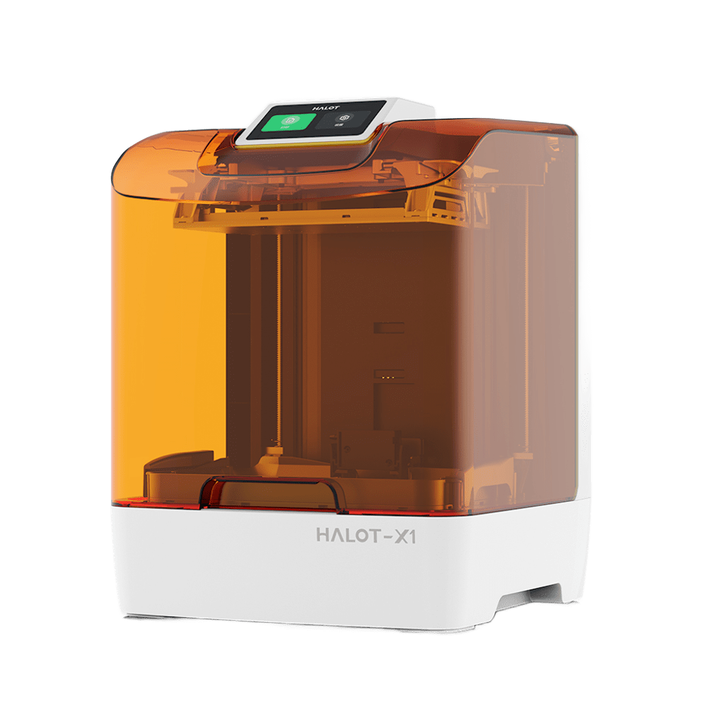
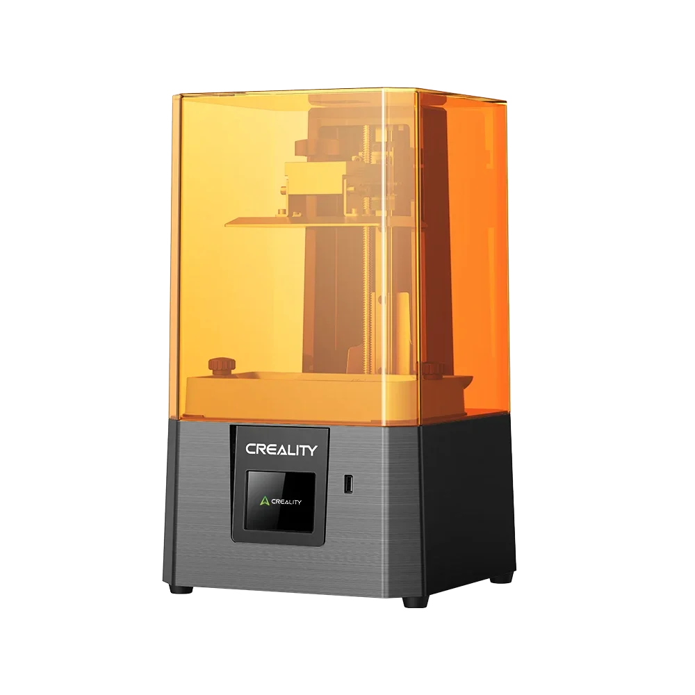
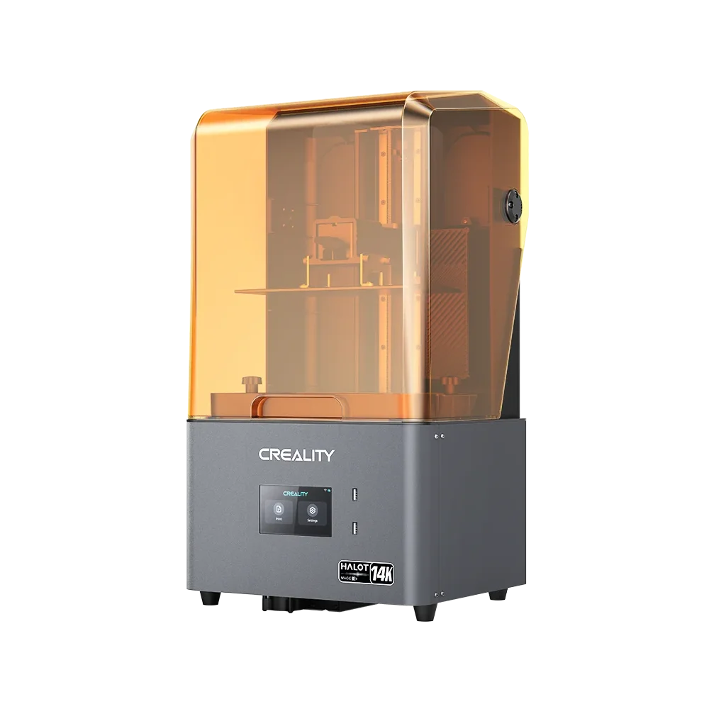

A surgeon holds a patient’s heart in their hands, studying each chamber, valve, and vessel before ever making a single incision. This is not hypothetical anymore. It’s happening in hospitals right now, using 3D-printed models designed from the patient’s own CT scan data. The same key technology is now sitting on desks in classrooms, makerspaces, and home offices globally.
3D heart print covers two very different worlds. One is clinical and life-saving. The other is educational, creative, and actually accessible to anyone with a 3D printer. This guide explores both how doctors are using 3D heart models to shift patient outcomes and exactly how you can print your own at home.
How Is 3D Printing Changing Heart Health?
The human heart is incredibly complicated. No two hearts are the same, and in patients with congenital heart disease, the differences between individuals can be dramatic. Classic imaging like CT and MRI scans show the heart in detail; however, they show it as a flat image on screen. A physical model changes that completely.
Here's how the process works in a clinical setting:
-
A patient’s heart is scanned through CT or MRI imaging
-
That imaging data is divided and can be exported into a series of 2 dimensional images. That is imported into software to produce 3D printed digital models
-
The model is sent to a 3D printer and created as a patient-specific physical replica
-
Surgeons, cardiologists, and trainees study, manage, and plan using the physical model
The clinical applications are verifiable and growing:
-
Congenital heart disease (CHD): Every child’s heart defect exhibits differently. According to a PMC research, 3D printed models notably enhance spatial understanding of complicated CHD anatomy for both trainees and clinicians. This exceeds what standard 2D and 3D imaging can convey.
-
Surgical planning: Surgeons practice intricate processes on a physical replica of the patient’s exact anatomy before entering the operating room. A 2021 review published in Frontiers in Cardiovascular Medicine verifies how 3D-printed models created in a low-cost clinical setting were used for surgical and interventional planning across 138 individual cases.
-
Patient and family communication: Surgeons use physical models to communicate with families and patients about exactly what’s wrong and what the procedure involves. The British Heart Foundation reports this practice at Great Ormond Street Hospital, where printed heart models are handed directly to families of children with complicated defects.
-
Medical education and training: Students and cardiac trainees use printed models to master 3D heart anatomy through touch, handling, and direct examination. Skills that conventional imaging and textbook diagrams simply cannot offer.
This is not experimental technology. Henry Ford Health's Center for Structural Heart Disease has produced over 1,000 3D-printed heart models for direct patient care since 2013, with three in-house printers dedicated entirely to this work. Hospitals around the world have built dedicated labs around this practice.
From Hospital 3D Printing Labs to Maker Desks
The healthcare industry and the maker world use a similar foundational procedure. A digital model gets sliced into layers and printed one layer at a time. The difference is in the starting point and the purpose.
Hospital labs start with patient imaging data, MRI or CT scans, modified through specialist slicing software into a patient-specific STL file. The outcome is a medically accurate replica of an individual’s heart, generated for a clinical purpose.
Students, makers, and educators work with pre-made anatomical model files, made for display, learning, education, or gifting. Such files are not patient-specific.
They are anatomically informed models that represent typical cardiac structure, designed for print testing and availability on a standard desktop printer.
The outcomes are different in accuracy and purpose. However, the physical thing- a detailed, manageable 3D heart anatomy model that presents chambers, vessels, and valves- is entirely possible on a consumer 3D printer at home.
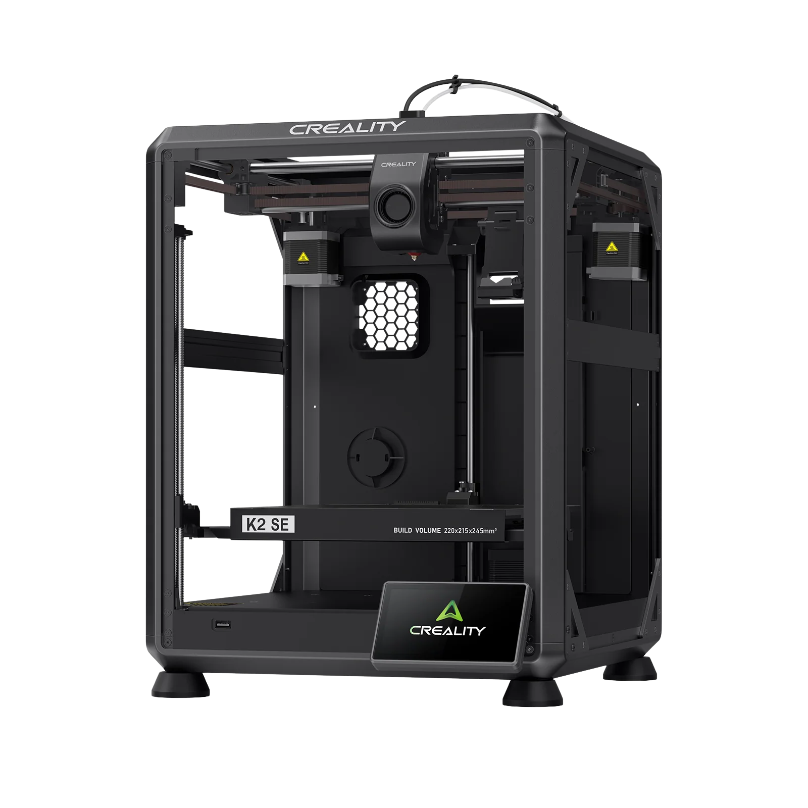
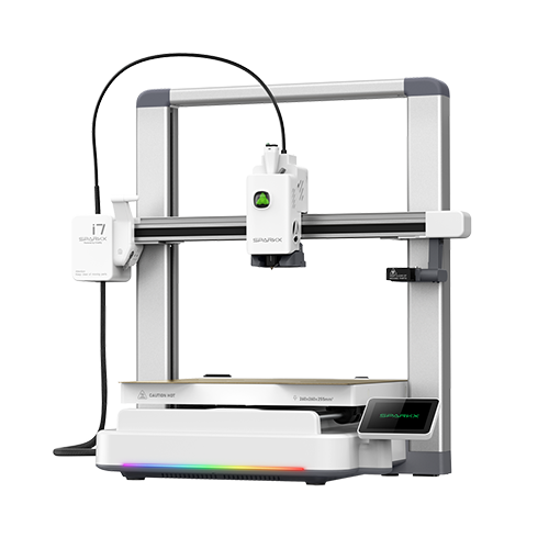
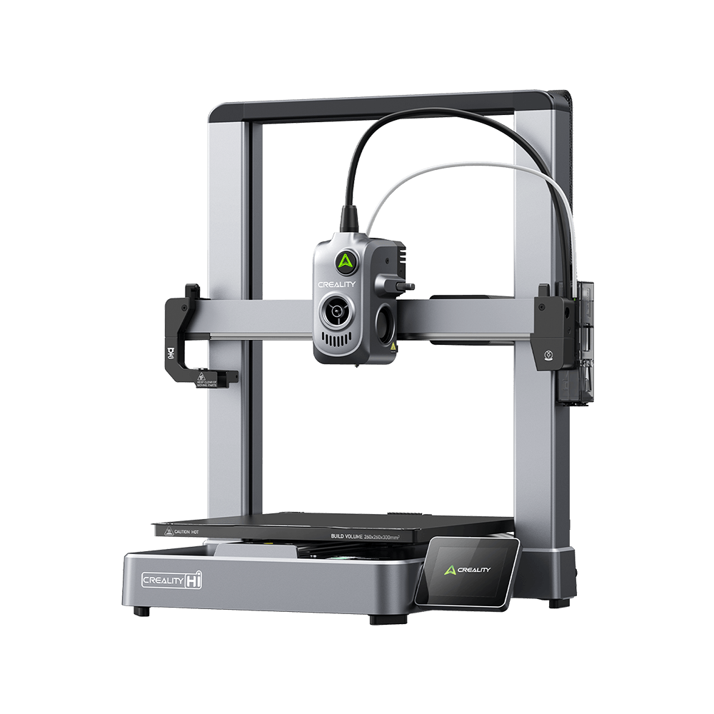
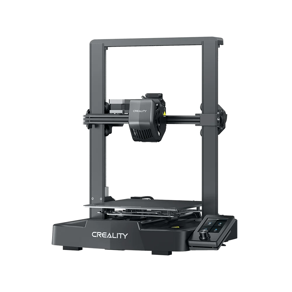
You don't need a hospital lab to print a heart model that teaches, displays, or impresses. You just need the right file, the right filament, and a printer that can handle the detail.
Where to Find 3D Printable Heart Models
The first decision isn’t about the printer or filament; it’s about the file. Heart 3D model files have several different categories, and selecting the correct one depends entirely on what you want to do with the finished print.
Anatomical Models
The most detailed choice: multi-part files that separate the heart into individual parts: atria, ventricles, valves, aorta, pulmonary vessels. Every component prints separately and assembles into a complete, accurate depiction of 3D heart anatomy. They take longer to print, anywhere from 10-24 hours.
Best for: biology students, anatomy enthusiasts, nursing and medical trainees, STEM classroom use.
Simplified Educational Models
Single-piece or two-part designs that reveal the heart’s overall shape and major features without the complexity of full anatomical separation. These print faster, require no assembly, and are more forgiving on print settings.
Best for: quick classroom demonstrations, beginner prints, introductory anatomy projects.
Decorative and Symbolic Hearts
Stylized heart forms, not anatomical, however visually striking. These extend from simple love-heart keepsakes to detailed fantasy designs. Print times are short and filament needs are minimal.
Best for: gifts, seasonal prints, Valentine's Day projects, desk decor.
Where to Find Heart 3D Model Files
Creality Cloud is the natural beginning point for Creality printer users. This is a model library with print-tested files from the community, accessible directly from the slicer. Moreover, you can discover free heart 3D model STL files on community platforms like Printables and Cults3D. They host both anatomical and decorative designs in varying levels of complexity and detail.
What Filament Works Best for a Heart Model?
Filament choice directly impacts a heart model’s feel, detail level, and educational value. Below is what works better for every use case:
-
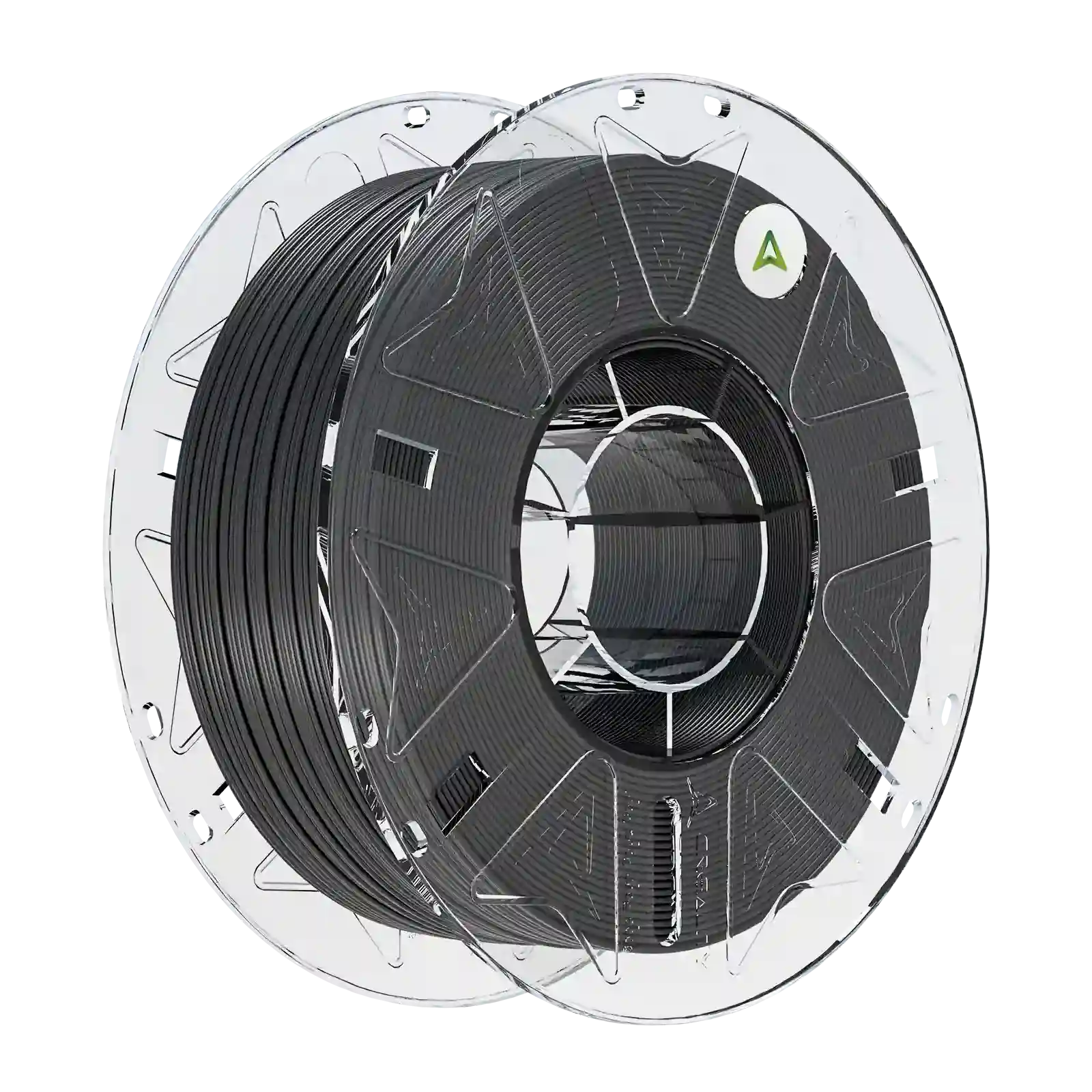
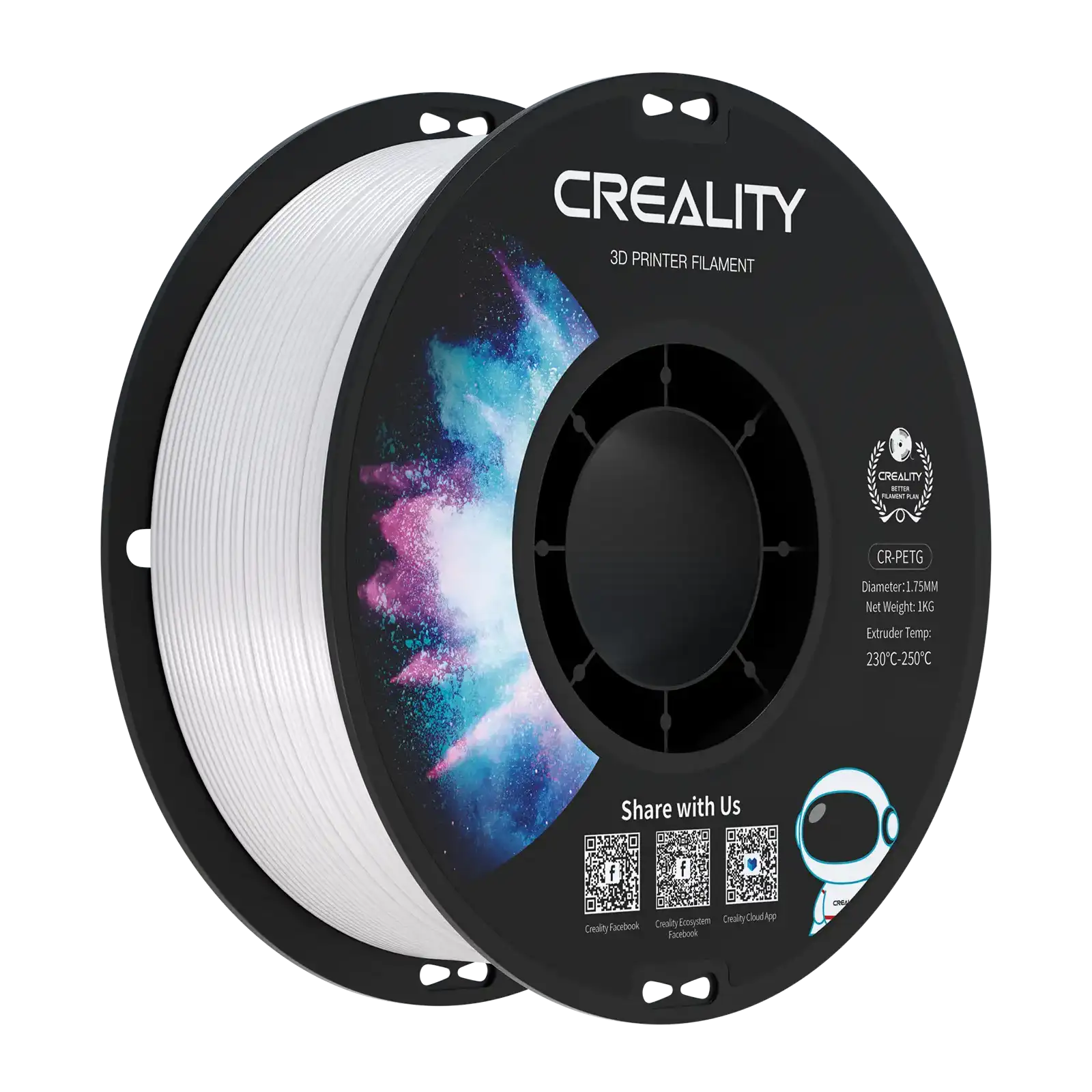
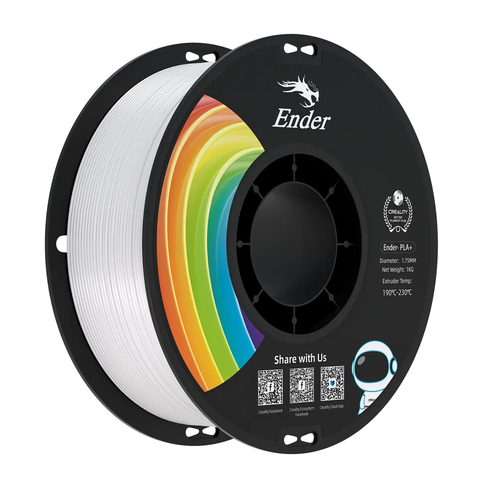
PLA: Easiest to print, readily available, has fine surface detail cleanly. Ideal for classroom display models and first-time anatomical prints. Rigid, so models hold their shape long-term.
-
TPU: Flexible filament that copies the soft, supple feel of actual tissue. Students can squeeze and analyze it, far closer to real anatomy than any stiff material. Needs slower speeds and a direct drive extruder
-
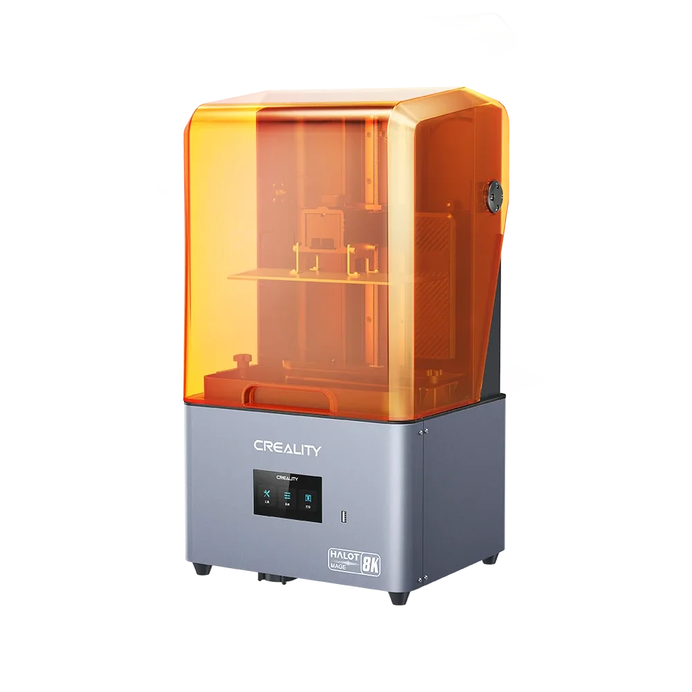
Resin: Generates the finest surface detail of any consumer printing process. Ideal for display-focused models where visual accuracy matters most. Less user-friendly, needs ventilation and post-processing.
-
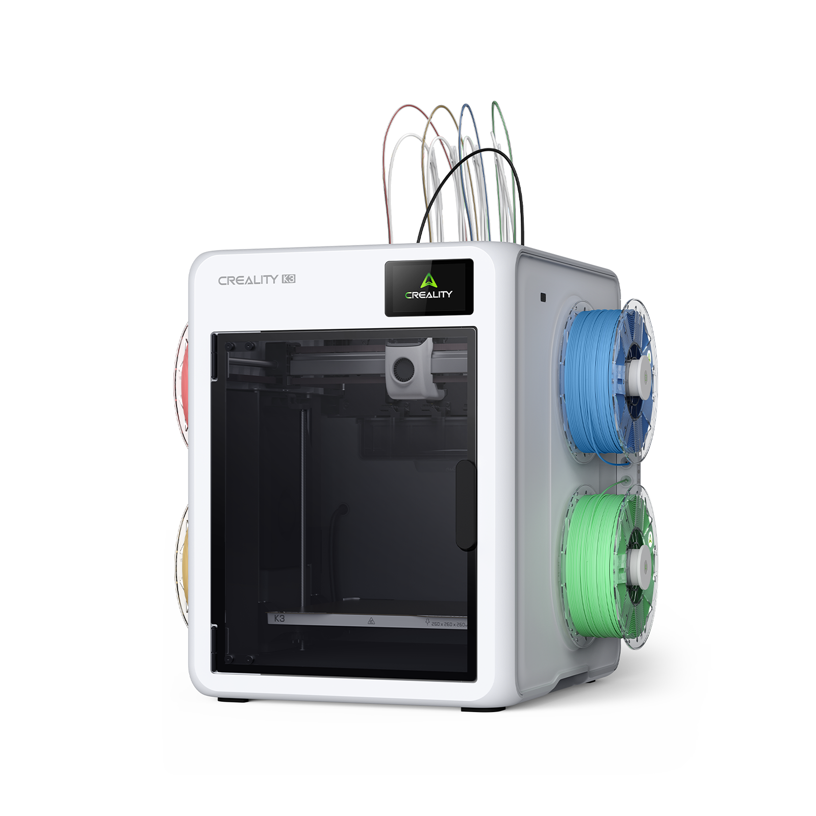
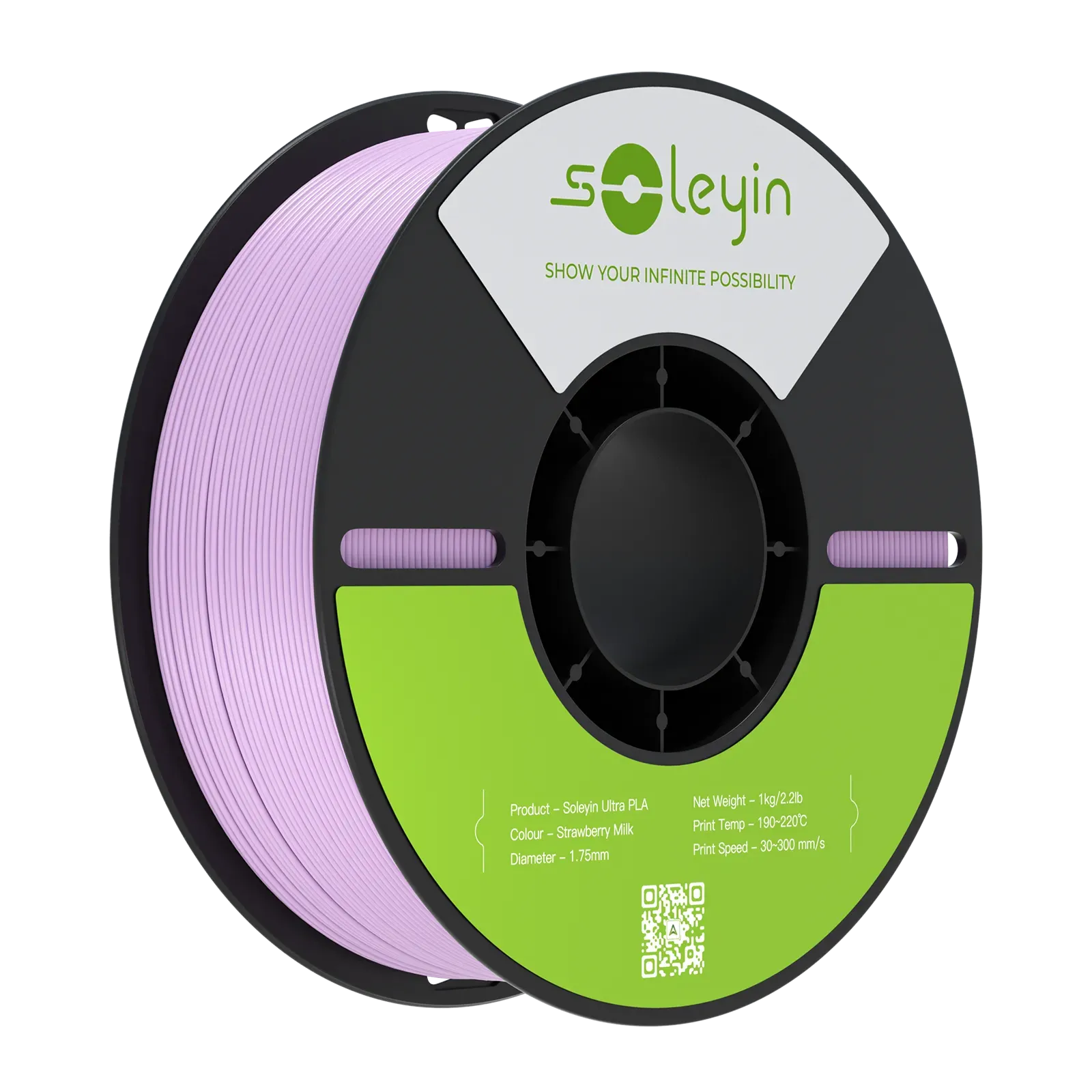
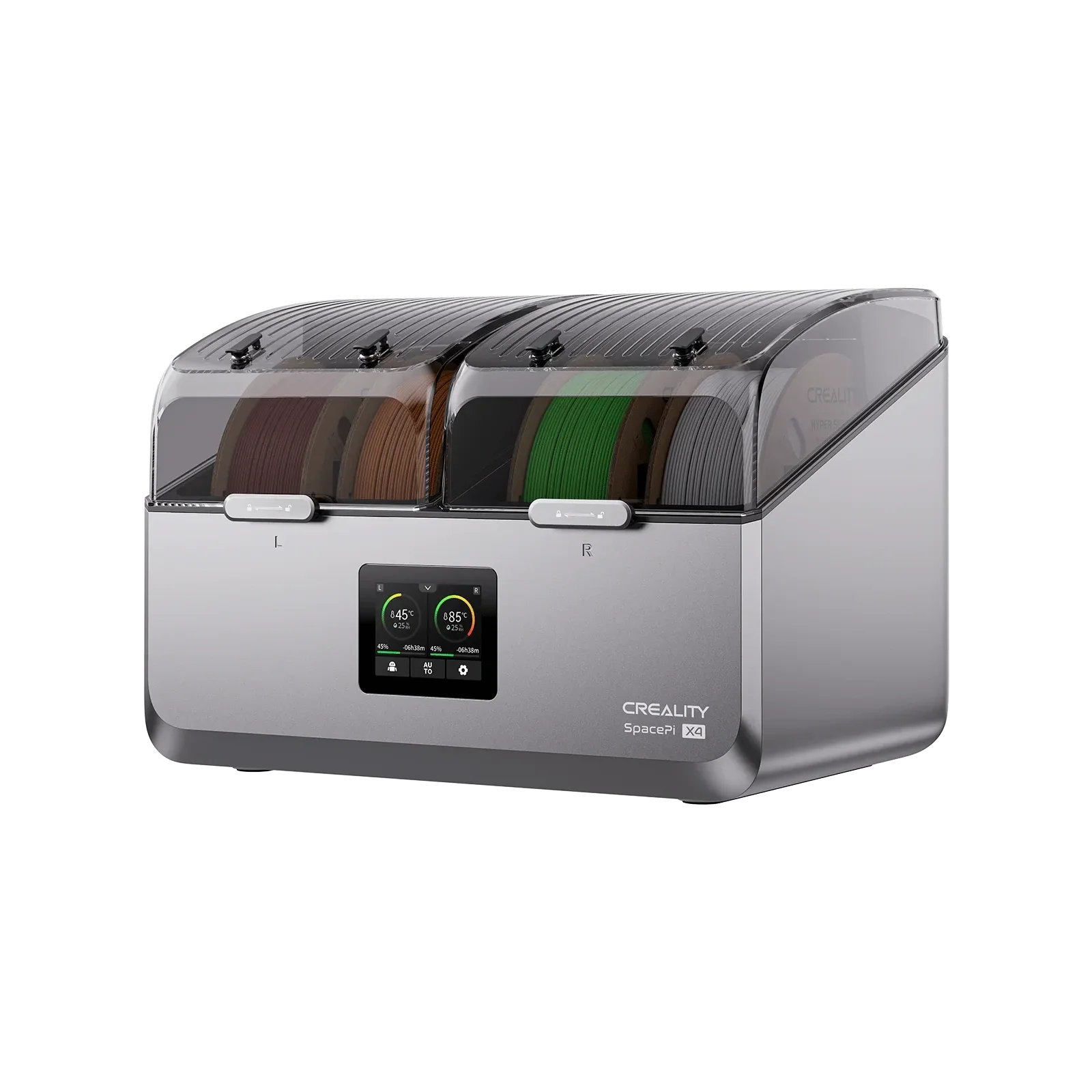
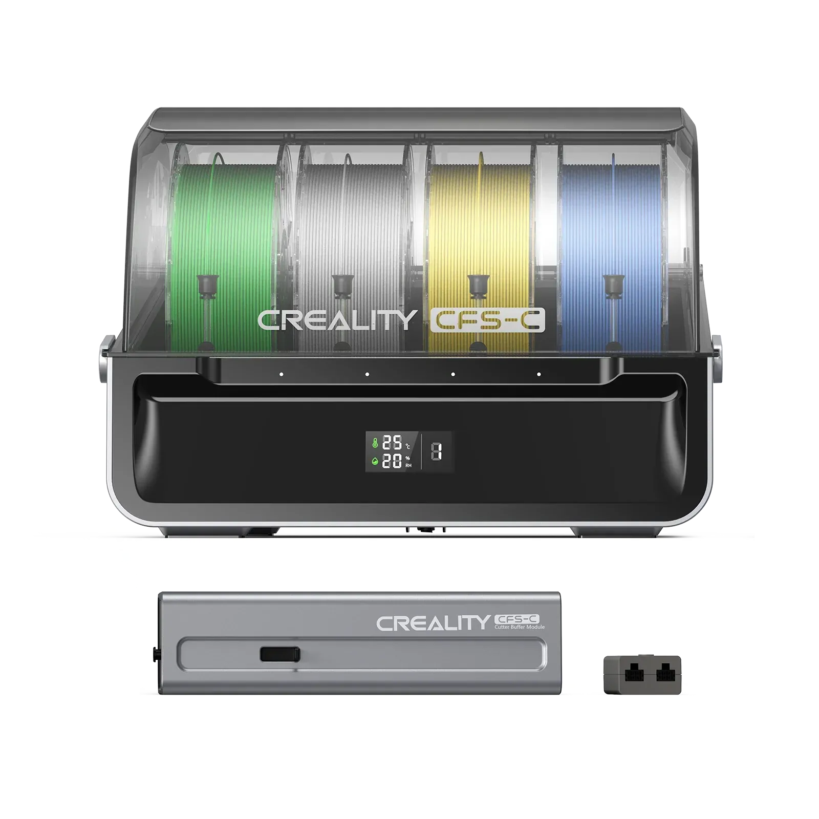
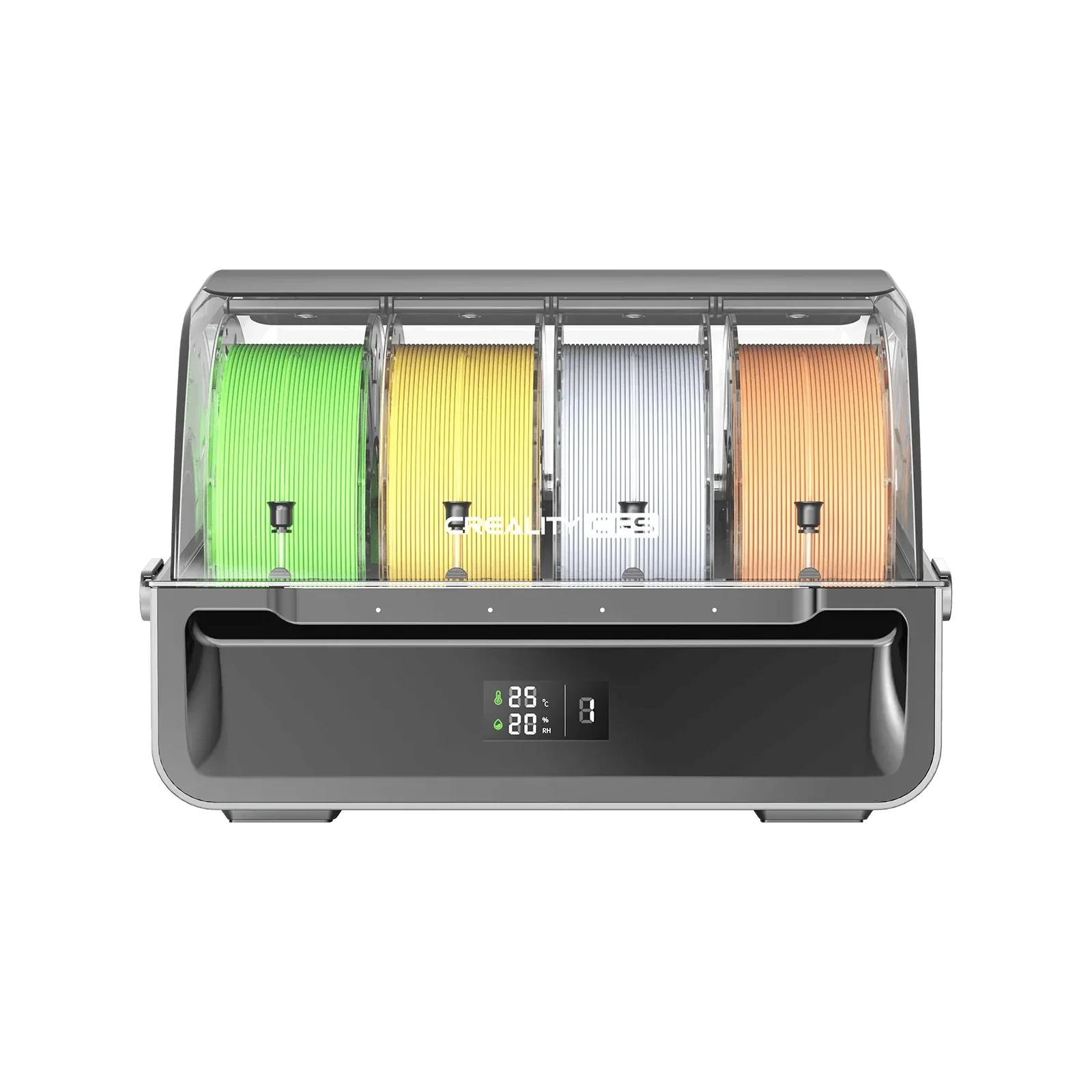
Multicolor PLA: Color-separating the anatomy (arteries in red, veins in blue, valve tissue in contrasting shades) converts a shape into an instructive tool. Creality’s Magic Box or 4-bay CFS manages color transitions automatically- no manual swaps.
|
Use Case
|
Recommended Filament
|
|
Classroom / educational display
|
PLA
|
|
Hands-on, tactile learning model
|
TPU
|
|
Multicolor anatomical accuracy
|
PLA with multicolor system (CFS / Magic Box)
|
|
High-detail display piece
|
Resin
|
Print Settings and Tips for Cleaner Results
Anatomical hearts require more from your printer than most standard models. These settings make the biggest difference:
-
Lower print speed by 20-30%: Fast travel moves produce a wavy surface on curved organic walls. Slower is cleaner.
-
Layer height: 0.21- 0.16 mm, finer than the default 0.2mm. The difference shows clearly on curved surfaces, valve edges, and vessel detail.
-
Use tree-style supports: on overhanging chamber structures. They’re easier to remove cleanly from detailed internal geometry than thick grid supports
-
Dry-fit multi-part models: before gluing; tolerances between separate ventricles, atria, and valves are tight. Test the fit first
Practical tip: First-layer adhesion matters more on long anatomical prints than almost any other model type. A failed first layer on a 10-hour heart print is the most common cause of wasted filament. Verify your Z-offset before starting.
Conclusion
The gap between a hospital 3D printing lab and a home printer is very small. Surgeons use 3D-printed hearts to plan life-saving operations. Students use them to understand anatomy, and makers use them to push their skills. Every result begins the same way: find the correct file, select the correct filament, and print. The free 3D heart model files are already out there. The printer settings are manageable. What's left is just deciding which version of this project is yours.